Smooth Muscle Diagram Labeled : Muscle The Histology Guide / It constitutes much of the musculature of
byAdmin-
0
Smooth Muscle Diagram Labeled : Muscle The Histology Guide / It constitutes much of the musculature of. See more ideas about muscle, skeletal muscle, histology slides. Smooth muscle anatomy smooth muscle tissue is also known as visceral muscle tissue. The given schematic diagram depicts heterodont teeth and its thecodont arrangement. Human anatomy muscle organ diagrams by julie ridge are part of my human anatomy series. The muscles of the human body can be categorized into a number of groups which include muscles relating to the head and neck, muscles of the torso or trunk, muscles of the upper limbs, and muscles of the lower limbs.
In skeletal muscle, a single type of somatic nervous system traverses to muscle, where it stimulates organelle in the muscle cells in order to release calcium. The majority of muscles in the leg are considered long muscles, in that they stretch great distances. Smooth muscle cells are found in the dividers of empty organs, including the stomach, digestion tracts, urinary bladder and uterus, and in the dividers of paths, for example, the supply routes and veins of the circulatory framework, and the tracts of the. The diagram is fully labeled. It is layered in a distinctive pattern of circular layers.
Perimysium Dense Irregular Connective Tissue Google Search Skeletal Muscle Muscle Cell Muscle Anatomy from i.pinimg.com Muscle fibers are organized into bundles supplied by blood vessels and innervated by motor neurons. In the body, there are three types of muscle: It is layered in a distinctive pattern of circular layers. See more ideas about muscle, skeletal muscle, histology slides. Individual muscle fibers, (b) surrounds groups of skeletal muscle fibers (fascicles), and (c) covers the muscle as a whole. Similar to skeletal muscle tissue, cardiac muscle does not regenerate to a great extent. The muscles of the human body can be categorized into a number of groups which include muscles relating to the head and neck, muscles of the torso or trunk, muscles of the upper limbs, and muscles of the lower limbs. Smooth muscle anatomy smooth muscle tissue is also known as visceral muscle tissue.
Individual muscle fibers, (b) surrounds groups of skeletal muscle fibers (fascicles), and (c) covers the muscle as a whole.
Relaxation of smooth muscle requires the myosin head to become dephosphorylated,. Smooth muscle makes up the walls of hollow organs, respiratory passageways, and blood vessels. Smooth muscle (textus muscularis levis) smooth muscle is a type of tissue found in the walls of hollow organs, such as the intestines, uterus and stomach. This smooth muscle can be found surrounding the walls of the blood vessels, the bronchioles in the lungs, and the sphincter muscles used in the gi tract.the gi tract, which is tubular by design, also houses longitudinal muscles in addition to the smooth. Give one difference between yudem and nh. Muscle fibers are organized into bundles supplied by blood vessels and innervated by motor neurons. Anatomy of a relaxed and contracted smooth muscle cell. See more ideas about muscle, skeletal muscle, histology slides. Smooth muscle is composed of sheets or strands of smooth muscle cells. Smooth muscle tissue, unlike striated muscle, contracts slowly and automatically. A gap junction (gj) is present in the longitudinal portion of the disk. The calcium is the cause of protein to detach from the actin and myosin fastly binds with the opening of actin. The smooth muscles perform the functions in the contrast of other types of muscles.
Structure and composition of muscle meat science. Kierszenbaum, al histology and cell biology 2nd ed., mosby elsevier, 2007, p. Skeletal muscles attach to and move bones by contracting and relaxing in response to voluntary messages from the nervous system. In this video i have shown the simplest way of drawing muscle drawing. Its wavelike movements propel things through the bodily system, such as food through.
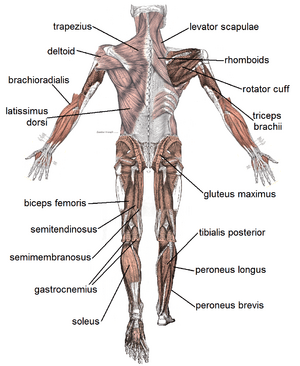
Muscular System Facts For Kids from kids.kiddle.co Individual muscle fibers, (b) surrounds groups of skeletal muscle fibers (fascicles), and (c) covers the muscle as a whole. Skeletal muscles attach to and move bones by contracting and relaxing in response to voluntary messages from the nervous system. Structure and composition of muscle meat science. Kierszenbaum, al histology and cell biology 2nd ed., mosby elsevier, 2007, p. Relaxation of smooth muscle requires the myosin head to become dephosphorylated,. Skeletal muscle tissue is composed of long cells called muscle fibers that have a striated appearance. Related posts of smooth muscle labelled diagram muscle anatomy shoulder. Full body muscles, both anterior and posterior diagrams;
Smooth muscle makes up the walls of hollow organs, respiratory passageways, and blood vessels.
1 response to simple smooth muscle diagram labeled unknown june 5, 2021 at 12:39 am. Smooth muscle tissue, unlike striated muscle, contracts slowly and automatically. It is layered in a distinctive pattern of circular layers. Structure and composition of muscle meat science. Skeletal muscles attach to and move bones by contracting and relaxing in response to voluntary messages from the nervous system. Smooth muscle is a type of muscle tissue which is used by various systems to apply pressure to vessels and organs. Smooth muscle tissue can regenerate from a type of stem cell called a pericyte, which is found in some small blood vessels. Muscle anatomy coloring sheets 12 photos of the muscle anatomy coloring sheets free muscle anatomy coloring sheets, muscle anatomy coloring pages, muscle anatomy coloring pages free, muscle anatomy coloring sheets, human muscles, free muscle anatomy coloring sheets, muscle anatomy coloring pages, muscle anatomy. It is the pen diagram of skeletal, smooth and cardiac muscle for. Give one difference between yudem and nh. Each slide has descriptions and images with a task for students to perform, such as labeling a diagram (based on the description) or. Smooth muscle cells are found in the dividers of empty organs, including the stomach, digestion tracts, urinary bladder and uterus, and in the dividers of paths, for example, the supply routes and veins of the circulatory framework, and the tracts of the. Intercalated disk (ventricle, cat) click to see enlarged view:
Related posts of smooth muscle diagram labeled muscle anatomy coloring sheets. Which type of muscle is labeled i in the venn diagram? In skeletal muscle, a single type of somatic nervous system traverses to muscle, where it stimulates organelle in the muscle cells in order to release calcium. Name the tough connective tissue cord that serves to attach a muscle to a bone. Smooth muscle anatomy smooth muscle tissue is also known as visceral muscle tissue.
In A Test Of Biology A Figure Of Smooth Muscle Labeled As A B C And D For Different Parts Of The Brainly In from hi-static.z-dn.net In skeletal muscle, a single type of somatic nervous system traverses to muscle, where it stimulates organelle in the muscle cells in order to release calcium. Kierszenbaum, al histology and cell biology 2nd ed., mosby elsevier, 2007, p. Intercalated disk (ventricle, cat) click to see enlarged view: Related posts of smooth muscle labelled diagram muscle anatomy shoulder. Muscle fibers are organized into bundles supplied by blood vessels and innervated by motor neurons. Diagram of smooth muscle cells. The given schematic diagram depicts heterodont teeth and its thecodont arrangement. Individual muscle fibers, (b) surrounds groups of skeletal muscle fibers (fascicles), and (c) covers the muscle as a whole.
This activity was created for distance learning during the 2020 pandemic as a substitution for traditional dissections and lessons that involve identifying the muscles and their function.
Smooth muscle is a type of muscle tissue which is used by various systems to apply pressure to vessels and organs. Smooth muscle cells are found in the dividers of empty organs, including the stomach, digestion tracts, urinary bladder and uterus, and in the dividers of paths, for example, the supply routes and veins of the circulatory framework, and the tracts of the. Similar to skeletal muscle tissue, cardiac muscle does not regenerate to a great extent. Its wavelike movements propel things through the bodily system, such as food through. Which type of muscle is labeled ii in the venn diagram? Pericytes allow smooth muscle cells to regenerate and repair much more readily than skeletal and cardiac muscle tissue. Smooth muscle anatomy smooth muscle tissue is also known as visceral muscle tissue. This smooth muscle can be found surrounding the walls of the blood vessels, the bronchioles in the lungs, and the sphincter muscles used in the gi tract.the gi tract, which is tubular by design, also houses longitudinal muscles in addition to the smooth. Muscle anatomy coloring sheets 12 photos of the muscle anatomy coloring sheets free muscle anatomy coloring sheets, muscle anatomy coloring pages, muscle anatomy coloring pages free, muscle anatomy coloring sheets, human muscles, free muscle anatomy coloring sheets, muscle anatomy coloring pages, muscle anatomy. Intercalated disk (ventricle, cat) click to see enlarged view: Skeletal (striated), smooth, and cardiac. It is the pen diagram of skeletal, smooth and cardiac muscle for class 10, 11 and 12. Smooth muscle tissue can regenerate from a type of stem cell called a pericyte, which is found in some small blood vessels.